Health
Science that holds the eye
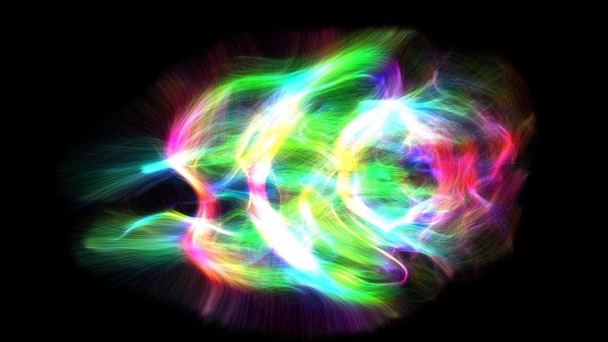
“Lightning of the Mouse Brain” by Alpen Ortug – “People’s Choice Award” winner. “The colors represent the directions of the nerve fibers. The two antenna-like structures on the left side of the image are the olfactory nerves, and the red region on the right side is the cerebellum,” said Ortug.
Alpen Ortug
You might not know what it is, but you’ll want to find out: ‘What story is this particular image trying to tell?’
Albert Einstein believed that mystery was the most beautiful experience humans could have. “It is the source of all true art and science,” he said.
But while scientists routinely probe nature’s mysteries, only rarely do they breach the divide into the world of art.
The Mass General Research Institute — home to more than 8,500 research scientists — is working to open a doorway between the two, hosting an annual contest to highlight outstanding scientific images. The Mass General Research Institute Image Awards were launched in 2018 to call attention to the research that underlies stunning images created through laboratory work and medical imaging. The 2023 winners were announced at a gala event last month. They include “Lightning of the Mouse Brain,” “The Arms Wide Open,” “A Living Puzzle,” “Fueling the Fight,” “Imaging the Brain,” and “It Takes Two.”
“Imaging the Brain” by Jennifer Guo – “Mixed Media” winner. “This illustration reflects my awe in learning how magnetic resonance imaging (MRI) is used to visualize the brain,” said Guo.
Jennifer Guo
“This is a great way to increase interest in science,” said Susan Slaugenhaupt, the institute’s scientific director and a professor of neurology at Harvard Medical School. “Though people don’t always know what they are, they’re beautiful images.”
Submissions for 2023 featured a swirl of colors and images. The gold, white, and turquoise of “Biological Fairy Lights” resemble the entangled chains of tiny outdoor lights but in fact are cells associated with cancer growth. More straightforward are the portraits of doctors, nurses, technicians, and reseachers that make up “Humans of MGRI.”
“A Living Puzzle” by Daniel Ruiz – “Science As Art” winner. A tumor biopsy from a patient diagnosed with head and neck cancer. Tumor cells are indicated in cyan.
Daniel Ruiz
“It Takes Two” by TJ Danenza – “Humans of MGRI” winner. Nurse Practitioner Madeline Macaluso (from left) and Facial Plastic Surgeon Linda Lee work together in the operating room. “This image visually emphasizes the critical role of teamwork and coordination in every surgical procedure,” said Danenza.
TJ Danenza
Slaugenhaupt noted that many of the pictures on first glance look like abstract art, drawing in viewers who then learn the stories behind the images.
“That story sometimes is as important as the image itself: Why did that submitter think this was a beautiful image? What story is this particular image trying to tell?”
“Fueling the Fight: Glutamine’s Critical Role in Brain Tumor Metabolism” by Sonu Subudhi – “A Closer Look” winner. “This image shows the expression of glutamine synthase by different brain structures and the accumulation of astrocytes in certain brain areas,” said Subudhi.
Sonu Subudhi
“The Arms Wide Open” by Bedri Karaismailoglu — “Across the Bridge” winner. “Standing tall with its two outstretched arms,” this image “represents an innovative, in-house-designed, and 3D-printed surgical guide meticulously engineered to provide precise and patient-specific solutions for ankle injuries,” said Karaismailoglu.
Bedri Karaismailoglu
More images
“The Crown of Telencephalon” by Alpen Ortug. A 3D representation of the white matter pathways in a human fetus at 14 weeks of gestation, as viewed from lateral (A) and top (B),” said Ortug.
Alpen Ortug
“A Simple Portrait” by TJ Danenza. “Linda Lee, MD, in the operating room.” Lee is a “member of the Mass Eye and Ear Facial and Cosmetic Surgery Center,” said Danenza.
TJ Danenza
“The Tree of Knowledge” by Sara Veiga. “A mouse dorsal root ganglion (DRG) was stained using a combination of four different fluorescently labeled antibodies,” said Veiga.
Sara Veiga
“Prototype Tangential Flow Device for Future Cancer Diagnostics” by Kilean Lucas and Joshua Spitzberg. “Using novel microfluidic chips containing a nanometer-scale porous membrane, we aim to filter and identify vesicles originating from early-stage tumors,” said Lucas and Spitzberg.
Kilean Lucas and Joshua Spitzberg
“Two Stage Facial Reanimation” by TJ Danenza. “Hoang Nguyen, MD, a PGY-2 in the Harvard Otolaryngology-Head and Neck Surgery residency program, harvests a muscle in the leg, which will be transferred into the face to aid in the facial reanimation of a patient with facial paralysis,” said Danenza.
TJ Danenza
“DWI-Based Retinotopic Mapping Prediction” by Nicolas Depauw. An image of “Retinotopy mapping, where each blob represents a brain hemisphere,” said Depauw.
Nicolas Depauw
“Broken Heart” by Eman Akam. “An image of the heart and the aorta of a mouse that underwent a procedure to mimic myocardial infarction, or a heart attack. Our molecular imaging with a fluorescent probe shows tissues in the aorta as well as the area of injury are abundant in a molecular target that is specific to wound healing,” said Akam.
Eman Akam, PhD
“Immune Surveillance of the DCIS” by Zuen Ren. “This picture shows the enrichment of immune cells in breast tissues including cytotoxic CD8 T cells (green) and natural killer (NK) cells (purple),” says Ren.
Zuen Ren


